
Respiratory System
Anatomy and Physiology Review – Respiratory System
Upper Respiratory Tract
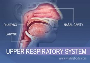
Nose
The respiratory cycle begins as air is inhaled through the nose, the nasal cavity warms, humidifies, and filters inhaled air (Thompson, 2018).
Oropharynx
The oropharynx is a hollow tube approximately 5 inches long; the structure is a passageway between the nose, sinuses, larynx, and trachea. Muscles of the oropharynx play a key role in speech and protect against the aspiration of food and fluids during swallowing (Thompson, 2018).
Lower Respiratory Tract
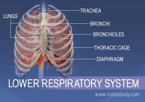
Trachea
The trachea comprises cartilage rings; these c-shaped rings extend from the bottom of the larynx to and behind the sternum. The trachea’s function allows air passage in and out of the lower respiratory tract (Thompson, 2018).
Bronchi
There are two bronchi, the right and the left. The right bronchus is smaller, wider, and more vertical than the left. The left bronchi, although smaller, is longer than the right. The right and left main bronchi function to warm and moisten air as it moves in and out of the respiratory tract (Thompson, 2018).
Bronchioles and Alveoli
The bronchioles are smaller branches of the mainstem bronchi that transport air to the alveoli. Alveoli are the smallest air sacs of the lungs. These tiny air sacs secrete surfactant, reducing surface tension and keeping the alveoli moist. Gas exchange of oxygen from the air breathed in and carbon dioxide circulating in the blood occurs at the alveoli level during each respiratory cycle (inhalation and exhalation). The PH of the blood (acid-base balance) is regulated based on the exchange of carbon dioxide (acid) and oxygen (base) in the alveoli during respiration. Changes in carbon dioxide levels influence the respiratory center in the brain by either speeding up or slowing down respiration to maintain a homeostatic acid-base balance (Thompson, 2018).
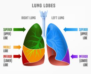
Lungs
The lungs are two cone-shaped air-filled structures. Like the left and right bronchi, the right lung is larger and heavier than the left and is divided into three separate lobes. The left lung is smaller and longer than the right and is divided into two lobes (Thompson, 2018).
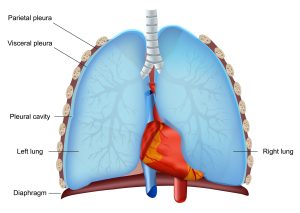
Pleura
Pleurae are two layered membranes that surround and protect the lungs. There are two types of pleurae: parietal and visceral. The parietal pleura lines and adheres to the thoracic wall, and the visceral pleura lines the outer surface of the lungs. The pleural cavity is the area between the parietal and visceral layers (Thompson, 2018).
Thoracic Cage
The thoracic or rib cage is a closed osseocartilaginous structure enclosing the thorax. It is formed by 12 thoracic vertebrae, 12 pairs of ribs, associated costal cartilages, and the sternum. This cavity protects and supports the upper body (Thompson, 2018; Bickley, 2021).
Diaphragm
The diaphragm is a dome-shaped muscle at the bottom of the chest cavity. The diaphragm is the primary muscle involved in respiration. The following video demonstrates how the diaphragm moves during inspiration and expiration (Bickley, 2021).
