Neurological System
Anatomy and Physiology Review – Neurological System
Central Nervous System
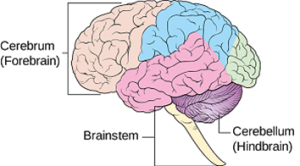
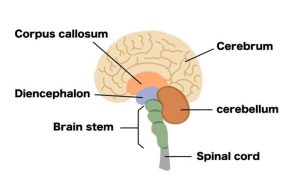
The brain is the largest part of the CNS; it contains gray and white matter and connective tissue called meninges that cover and protect the brain and spinal cord. The brain is made up of three parts: the cerebrum, cerebellum, and brainstem (Thompson, 2018).
Cerebrum
The Cerebrum is the largest part of the brain; it is divided into the right and left hemispheres and separated by a longitudinal fissure called the corpus callosum. The corpus callosum allows two-way communication between the hemispheres. These cerebral hemispheres are further divided into four areas known as the frontal, parietal, temporal, and occipital lobes (Thompson, 2018).
Cerebellum
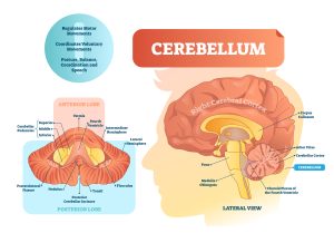
The cerebellum is in the back of the brain. It links sensory input from CNS pathways (sensory and motor) with movement, coordination, speech, and the five senses of sight, hearing, smell, touch, and taste. If the cerebellum becomes damaged, a person may experience problems with balance and coordination (Thompson, 2018).
Brainstem
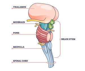
The brainstem is located in the posterior portion of the brain; it connects the cerebrum with the spinal cord and transmits impulses from the spinal cord to the brain. The brainstem controls involuntary behaviours such as breathing, heart rate, and sleep and comprises three vital structures, each carrying out a specific relay function. The medulla oblongata (bottom portion of the brainstem) coordinates head and eye movement. It houses core neurological centers that control involuntary bodily processes such as blood pressure, heart rate, breathing, and digestion. The pons (middle portion of the brainstem) relays messages between the cerebrum and cerebellum and is primarily responsible for sensory and motor function. The midbrain (top of the brainstem) is often described as the brain’s “information highway,” coordinating sensory and motor information of the eyes, ears, facial sensation, and upper limbs (Thompson, 2018).
Central Nervous System – Two Main Pathways
The CNS has two main pathways:
Ascending Pathways
Ascending pathways for transmission of sensory data such as pain up the spinal cord to the brain. Sensory data is relayed through the spinothalamic tract and posterior columns.
- Spinothalamic tract – a pathway that relays sensory impulses to the brain registering peripheral sensations of pain, temperature, and crude touch
- Posterior columns – transmit peripheral sensations of vibrations, proprioception, kinesthesia, pressure, and fine touch.
Descending Pathways
Descending pathways for transmission of motor data, such as moving one’s arm, from the brain down the spinal cord to the body. Motor system data is carried via the corticospinal tract, basal ganglia, and cerebellar systems.
- Corticospinal (pyramidal) tract – the primary motor system that controls voluntary movement, consists of both upper and lower motor neurons
- Basal Ganglia system – helps to maintain normal muscle tone and body movement such as walking
- Cerebellar system – primarily responsible for coordinating motor activity, maintaining equilibrium, and posture.
Peripheral Nervous System
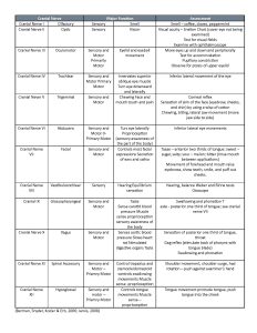
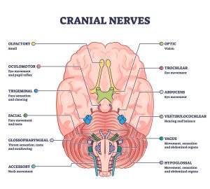
The PNS comprises 12 cranial nerves, 31 pairs of spinal nerves, and the autonomic nervous system’s two divisions (sympathetic and parasympathetic).
PRINTABLE 3.0
Spinal Cord and Nerves
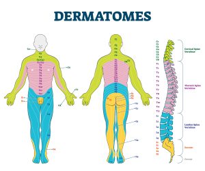
The spinal cord is the primary connection between the brain and the body. There are eight cervical pairs, twelve thoracic, five lumbar, five sacral, and one coccygeal pair. Each pair is named after the spinal vertebrae just below the nerve exit point along the spinal column. Each nerve is attached to the spinal cord by two nerve roots: sensory (afferent) and motor (efferent). The sensory root of each spinal nerve innervates with an area of the body’s skin surface called a dermatome (Thompson, 2018).
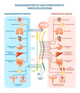
Autonomic Nervous System
The part of the peripheral nervous system that regulates involuntary physiological processes such as the beating of the heart, the process of digestion, and even sexual arousal. The two main systems within the autonomic nervous system are the parasympathetic nervous system (PNS), or “rest and digest,” and the sympathetic nervous system (SNS), also known as the “fight or flight” (Thompson, 2018).