Female Genitalia and Rectal System
Anatomy and Physiology Review – Female Genitalia and Rectal System
Female Reproductive


External Genitalia
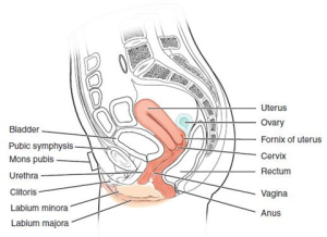
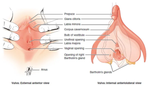
The external female reproductive structures are collectively referred to as the vulva and include the mons pubis, labia majora and minora, clitoris, urinary meatus, Bartholin glands, and the opening to the vagina (introitus ).

Internal Reproductive Organs
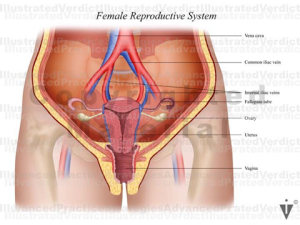
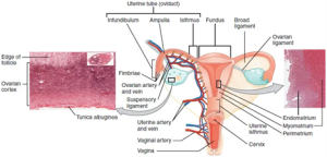
Internal reproductive organs include the vagina, uterus, cervix, fallopian tubes, and ovaries.
- The vagina is a hollow and expandable muscular canal that extends from the external genitalia to the cervix.
- The uterus is a pear-shaped muscular organ between the urinary bladder and the rectum. It consists of two parts: the fundus, located at the top of the uterus and the isthmus, the lower portion of the uterus. The position of the uterus in the female pelvis can be anteverted (tilted toward the abdomen), retroverted (tilted backwards toward the rectum), or midline (Betts et al., 2022; Thompson, 2018).
- The cervix is part of the lower uterus and is a canal that secretes mucous. The opening of the cervix is called the external os and is in the vagina.
- Fallopian tubes (uterine tubes) are tube-like structures measuring 10-12 cm. The tubes extend from the left and right sides of the fundus and branch out close to each ovary. The tubes use ciliary and muscular waves to move a mature egg toward the uterus (Betts et al., 2022; Thompson, 2018).
- Ovaries are almond-shaped organs measuring 3-4 cm in length and 2 cm in width, located on each side of the uterus. The ovaries produce the female hormones of estrogen and progesterone and contain female eggs (Betts et al., 2022; Thompson, 2018).


