Head, Eyes, Ears, Nose and Throat System
Anatomy and Physiology Review – Head, Eyes, Ears, Nose and Throat System
Head
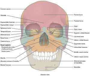
The head comprises two specific parts: the face and the cranium (skull). There are 22 bones in the skull, and all structures of the face and skull are connected by immovable joints called sutures and connective tissue. The anterior portion of the skull makes up the face and consists of 14 facial bones. The temporal artery provides blood supply to the head (Betts et al., 2022).
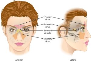
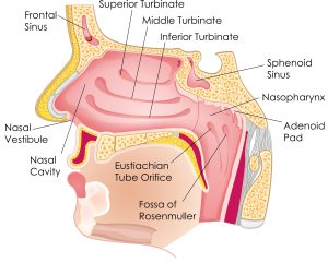
The face houses the sinuses, air-filled cavities that reduce the overall weight of the skull and provide voice resonance (Thompson, 2022). There are four pairs of sinuses in the face: frontal, above the eyes (palpable); ethmoid, between the eyes deep in the skull (non-palpable); sphenoid, behind the nasal cavity (non-palpable); and maxillary, in cheekbones below the eyes (palpable).
Eyes
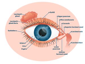
The internal and external structures of the eye work together to support human sight. Three cranial nerves are involved in vision (CN III, IV, VI). The perception of sight begins as light waves that travel from the cornea to the retina, where they are transformed into nerve impulses that travel to the brain via the optic nerve.
Extraocular structures include the eyebrows, eyelashes, eyelids, and lacrimal glands. The Intraocular structures include extraocular muscles, aqueous humor, choroid, iris, lens, pupil, posterior chamber, cornea, fundus, macula, optic disc, and the retina and its accompanying blood vessel structure (Betts et al., 2018; Thompson, 2018).
Ears
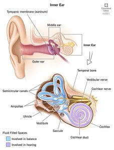
The ear is essentially a three-part structure. The first part is the outer ear and the ear canal. The second is the middle ear, located directly behind the tympanic membrane. Three small bones are in this area: the malleus, the incus, and the stapes. The third part is known as the inner ear. This part of the ear functions to control hearing and balance. The inner ear structures include the cochlea, semi-circular canals (labyrinth), and the vestibule. The third part is the inner ear. Each part of the ear has a specific function: the outer ear collects, and channels sound into the ear canal; the middle ear separates the ear canal from the inner ear and ensures that adequate amounts of sound energy are picked up by the fluid in the cochlea. The inner ear translates sound energy into recognizable information and regulates balance (Betts et al., 2022).
Nose
The nose comprises bone and cartilage and is physiologically part of the upper respiratory system. Internal structures of the nose include the nares, the nasal septum, the turbinates, and the adenoids. The turbinates are mucous membrane-covered bone structures, and the adenoids are clusters of lymphatic tissue (Thompson, 2018).
Throat
Mouth
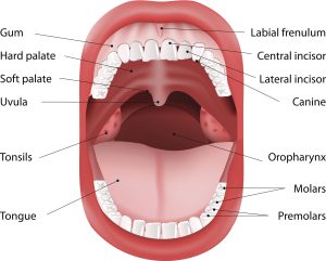
The mouth is covered by a mucous membrane (oral and buccal mucosa), and the lips separate the external mouth from the internal oral cavity. The oral cavity houses the hard and soft palates, the mandible, Stenson’s ducts (parotid duct), and Wharton’s ducts. Stenson’s ducts allow saliva to flow from the parotid gland into the mouth, whereas Wharton’s ducts transport saliva from the submandibular glands and drain saliva produced by the sublingual glands. There are 32 pairs of teeth in the adult mouth rooted in the gingiva (gums) and the tongue, a muscle located on the floor of the mouth (Thompson, 2018).
Pharynx
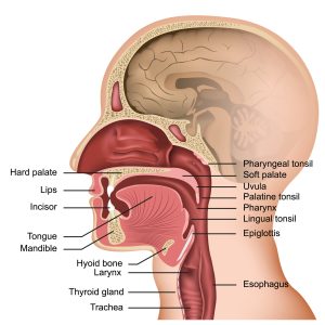
The pharynx is divided into three regions: the nasopharynx, oropharynx, and laryngopharynx. Below the pharynx are the trachea, epiglottis, and tonsils. The trachea is a tube composed of cartilage and membranes; the average adult trachea measures approx. four inches in length and ¾ to one inch in diameter. The epiglottis is a flap of tissue that primarily separates the trachea and the esophagus to prevent aspiration of food and fluid. Tonsils are part of the immune system, composed of lymphatic tissue and sit at the back of the pharynx (Betts et al., 2022).
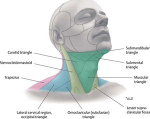
Neck
The neck contains many vital structures, including seven cervical vertebrae, the sternocleidomastoid and trapezius muscles, the thyroid gland, numerous cervical lymph node chains, the trachea, and the carotid arteries. For descriptive purposes, the anterior and lateral necks are divided into two triangles, which share the sternocleidomastoid muscle (SCM) as a boundary. Each triangle is further divided into smaller triangles (Thompson, 2018).
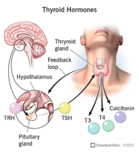
The thyroid gland is shaped like a butterfly, with two lobes four to six cm long. The thyroid isthmus joins the two lobes together. The thyroid gland releases several hormones, most notably TSH (thyroid stimulating hormone), T3 (triiodothyronine), and T4 (thyroxine). T3 and T4 hormone release is controlled by a feedback loop initiated by the pituitary gland based on TSH levels; when T# and T4 levels increase, less TSH is produced and vice versa (Cleveland Clinic, 2021 ). Thyroid hormones regulate body metabolism, temperature, heart rate, and muscle contraction (Thompson, 2018).