Musculoskeletal System
Anatomy and Physiology Review – Musculoskeletal System
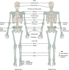
The skeleton consists of 206 bones and is divided into two regions for easy classification. The two regions are the axial skeleton and the appendicular skeleton.
Skeleton
Axial Skelton
The axial skeleton is composed of the head, vertebrae, ribs, and sternum.
Appendicular skeleton
The Appendicular skeleton comprises 126 bones, including the upper and lower limbs, the pelvis, clavicles, and scapulae. The bones that attach each upper limb to the axial skeleton form the pectoral girdle (shoulder girdle). The pectoral girdle consists of the scapula and clavicle. The bones that attach the lower limbs to the axial skeleton form the pelvic girdle. The pelvic girdle consists of one single bone called the hip bone. Each hip bone attaches to the other to form the bony pelvis, which consists of the hips, the sacrum, and the coccyx (Betts et al., 2022).

Classification of Bones
Bones are dense connective tissue providing structure, protection, and mobility. Bones are classified as long bones or short irregular bones. This classification is based on their size and structure (Thompson, 2018).
Long Bones
The long bones provide the structure for the arms and legs, including the humerus, ulna, radius, femur, tibia, and fibula. These long bones may also produce bone marrow and blood cells (Thompson, 2018).
Irregular Short Bones
Irregular or short bones provide structure for the ankles, feet, wrists, and hands.
The upper limbs are divided into three regions: the arm, the forearm, and the hand. The lower limbs are divided into the thigh, leg, and foot (Betts et al., 2022).
Upper Limbs
Humerus and Elbow Joint
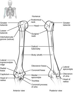
The humerus is the single bone of the upper arm region. It articulates with the radius and ulna bones of the forearm to form the elbow joint (Betts et al., 2022).
Ulna and Radius
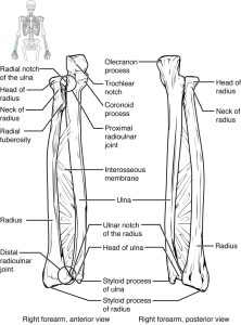
The ulna is located on the medial side of the forearm, and the radius is on the lateral side. An interosseous membrane attaches these bones (Betts et al., 2022).
Bones of the Wrist and Hand
The eight carpal bones form the base of the hand. These are arranged into proximal and distal rows of four bones each. The metacarpal bones form the palm of the hand. The thumb and fingers consist of the phalanx bones (Betts et al., 2022).
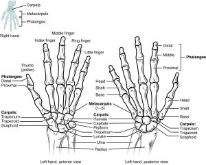
Lower Limbs
Pelvis
A single hip bone forms the pelvic girdle. The hip bone attaches the lower limb to the axial skeleton through its articulation with the sacrum. The right and left hip bones, the sacrum, and the coccyx form the pelvis (Thompson, 2018).
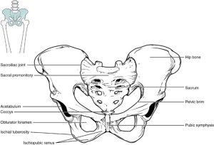
The Hip Bone
The adult hip bone consists of three regions. The ilium forms the large, fan-shaped superior portion, the ischium forms the posteroinferior portion, and the pubis forms the anteromedial portion (Betts et al., 2022; Bickley, 2021).
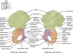
Femur and Patella
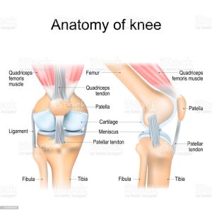
The femur is the single bone of the thigh region. It articulates superiorly with the hip bone at the hip joint and inferiorly with the tibia at the knee joint. The patella only articulates with the distal end of the femur (Betts et al., 2022; Bickley, 2021).
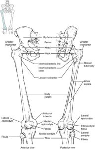
Tibia and Fibula
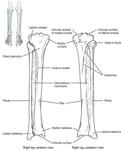
The tibia is the larger, weight-bearing bone on the leg’s medial side. The fibula is the slender bone of the lateral side of the leg and does not bear weight (Betts et al., 2022).
Bones of the Foot
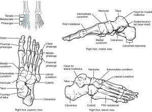
The bones of the foot are divided into three groups. The seven tarsal bones form the posterior foot. The mid-foot has five metatarsal bones. The toes contain phalanges (Betts et al., 2022).
Joints
A joint is where two or more bones come together; some joints are movable, and some are not. Joints that move allow functional movement of body parts. Ligaments often stabilize joints and may have tendons that cross the joint to allow movement. Many joints, all moving in unison, create opportunities for the skeletal system to move seamlessly in many directions. The two main classifications of joints are synovial and non-synovial (Betts et al., 2022; Bickley, 2021).
Synovial Joints
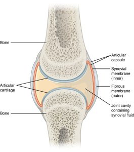
Synovial joints allow for smooth movements between the adjacent bones. The joint is surrounded by an articular capsule that defines a joint cavity filled with synovial fluid. A thin layer of articular cartilage covers the articulating surfaces of the bones. Ligaments support the joint by holding the bones together and resisting excess or abnormal joint motions (Betts et al., 2022; Bickley, 2021).
Types of Synovial Joints
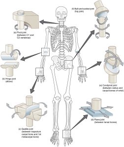
The six synovial joints allow the body to move in various ways. (a) Pivot joints allow for rotation around an axis, such as between the first and second cervical vertebrae, which allows for side-to-side rotation of the head. (b) The hinge joint of the elbow works like a door hinge. (c) A saddle joint is the articulation between the trapezium carpal bone and the first metacarpal bone at the base of the thumb. (d) Plane joints, such as those between the tarsal bones of the foot, allow for limited gliding movements between bones. (e) The radiocarpal joint of the wrist is a condyloid joint. (f) The hip and shoulder joints are the only ball-and-socket joints of the body (Betts et al., 2022; Thompson, 2018).
VIDEO 1.2
Non-synovial Joints consist of fibrous and cartilaginous joints.
Fibrous Joints
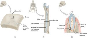
Fibrous Joints form strong connections between bones. (a) Sutures join most bones of the skull. (b) An interosseous membrane forms a syndesmosis between the radius and ulna bones of the forearm. (c) A gomphosis is a specialized fibrous joint that anchors a tooth to its socket in the jaw (Thompson, 2018).
Cartilaginous Joints
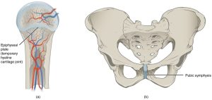
Cartilaginous joints bones are united by hyaline cartilage to form synchondrosis or fibrocartilage to form a symphysis. (a) The hyaline cartilage of the epiphyseal plate (growth plate) forms a synchondrosis that unites the shaft (diaphysis) and end (epiphysis) of a long bone and allows the bone to grow in length. (b) The pubic portions of the pelvis’s right and left hip bones are joined by fibrocartilage, forming the pubic symphysis (Betts et al., 2022).
Vertebrae
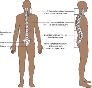
The vertebral column, also known as the spine column, is composed of 24 vertebrae (each separated by a vertebral disc), plus the sacrum and the coccyx. This column supports the head, neck, and body and protects the spinal cord housed within the vertebral column. The vertebrae are divided into three regions: cervical C1–C7 vertebrae, thoracic T1–T12 vertebrae, and lumbar L1–L5 vertebrae (Betts et al., 2022; Thompson, 2018). The vertebral column has two primary curvatures (thoracic and sacrococcygeal curves) and two secondary curvatures (cervical and lumbar).
Muscles
There are three primary classifications of muscle tissue: Skeletal muscle is attached to bones, and its contraction makes possible locomotion, facial expressions, posture, and other voluntary movements of the body. Cardiac muscle forms the contractile walls of the heart. Smooth muscle tissue contraction is responsible for involuntary movements in the internal organs. It forms the contractile component of the digestive, urinary, and reproductive systems, airways, and arteries (Betts et al., 2022).
| Tissue | Histology | Function | Location |
| Skeletal | Long cylindrical fibre, striated, many peripherally located nuclei | Voluntary movement produces heat, protects organs | Attached to bones and around entrance points to the body (e.g., mouth, anus) |
| Cardiac | Short, branched, striated, single central nucleus | Contracts to pump blood | Heart |
| Smooth | Short, spindle-shaped, no evident striation, single nucleus in each fibre | Involuntary movement moves food, involuntary control of respiration, moves secretions, regulates the flow of blood in arteries by contraction | Walls of major organs and passageways |
Muscle Strength Scale
The movements of joints are described and referenced from an anatomically neutral position.
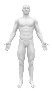
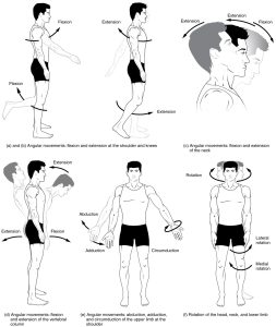
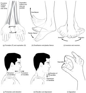
There are a variety of joint movements that can occur. Abduction is the movement away from the midline, and adduction is the movement toward the body’s midline. Pronation is inward rotation away from the neutral position, and supination is a rotational movement toward the neutral position. Eversion moves away from the body’s midline, and eversion moves toward the body’s midline. Dorsiflexion is the upward movement of the foot, and plantar flexion is the downward movement of the foot. Flexion is the movement of a joint toward the body; extension is away from the body, and elevation is an upward movement of a body part. These terms become important when documenting joints’ range of motion (ROM) during an MSK physical exam (Betts et al., 2022; Bickley, 2021).
Tendons and Ligaments
Tendons are made of dense connective tissue; this connective tissue is how muscles and bone are connected. If a tendon injury occurs, it is called a strain. Ligaments attach bone to bone and are non-elastic. Injury to a ligament is called a sprain. Both ligaments and tendons provide support to joints (Betts et al., 2022).
