Integumentary System
Anatomy and Physiology Review – Integumentary System
Skin
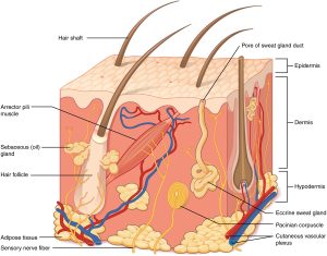
The skin is divided into two layers: the epidermis, which is made up of tightly packed epithelial cells, and the dermis, which is made up of dense, irregular connective tissue that houses blood vessels, hair follicles, sweat glands, and other structures. The hypodermis lies beneath the dermis and is composed primarily of loose connective and fatty tissues (Betts, 2022).

Hair
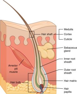
Hair follicles begin in the epidermis and have numerous parts. The medulla is the central core of the hair, surrounded by the cortex, a layer of compressed, keratinized cells covered by an outer layer of very hard, epithelial cells known as the cuticle. Hair texture (straight or curly) is determined by the shape and structure of the cortex (Betts, 2022).

Nails
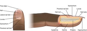
Nails are accessory structures of the integumentary system. The nail bed is a specialized epidermis structure found at the tips of our fingers and toes. The nail body is formed on the nail bed and protects the tips of our fingers and toes because they are the body’s farthest extremities and the parts that experience the most mechanical stress. Additionally, the nail body functions as a back support for picking up small objects with the fingers (Hesse et al., 2017).


