
Male Genitalia and Rectal System
Anatomy and Physiology Review – Male Genitalia and Rectal System
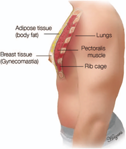
Breasts
Male breast tissue includes fibrous, glandular, and fatty tissue, the breasts do not enlarge because of the male hormone testosterone.
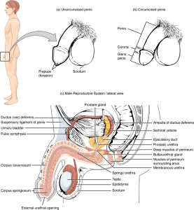
Penis
The penis is divided into three sections: the radix (or root), which is attached to the abdominal wall; the shaft (or body); and the glans penis (or head of the penis). The male urethra is situated on the tip of the penis.
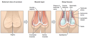
Scrotum
The scrotum envelopes the testes in a muscular sack that extends behind the shaft of the penis. The scrotum is divided into two compartments by the dartos muscle; each compartment holds one testis. Two cremaster muscles extend from the abdominal wall to cover each teste in a netlike fashion.
Testes
The testicles are male reproductive organs that produce sperm and testosterone. These paired oval-shaped organs are housed in the scrotum and protected in a double layer of connective tissue. Testes are divided into hundreds of lobules where the male sperm develops in seminiferous tubules (Betts et al., 2018).
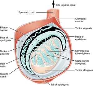
Epididymis
The epididymides are located above and behind each teste; their primary role is to collect and hold mature sperm. The physical structure of the epididymides is that of a long coil (if placed in a straight line, it would be approximately six meters long). Sperm travels through these coils and matures along the way. It can take up to twelve days for the sperm to pass through the epididymides (Thompson, 2018).
The Testicular Duct System
The testicular duct system comprises the vas deferens and the spermatic cord. During ejaculation, the sperm are pushed to the vas deferens, a muscular tube enclosed and supported by the spermatic cord (Betts et al., 2022).
Seminal Vesicles
Glands that add volume to the sperm to make seminal fluid. The fluid the seminal vesicles contribute is high in fructose, which the sperm uses to promote the formation of ATP to support sperm movement further. The newly produced seminal fluid moves to the ejaculatory duct, transporting it to the prostate gland (Betts et al., 2022).
Prostate Gland
The prostate gland sits between the bladder and the rectum and is about the size of a walnut. This gland receives the seminal fluid-now called semen and excretes an alkaline substance into the semen that allows coagulation and de-coagulation of the semen once ejaculated. Once in the female vagina, the thickness of the semen dissipates, and the semen returns to a fluid state which can quickly move deeper into the female reproductive tract (Thompson, 2023).
