Abdominal System
Anatomy and Physiology Review – Abdominal System
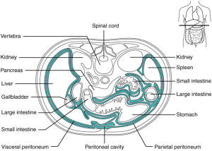
The abdomen is the largest cavity in the human body; it houses the primary digestive organs and is covered by an endothelial serous membrane lining called the peritoneum. There are two types of peritoneal linings in the abdominal cavity: the first type of lining covers the walls of the abdominal cavity and is called the parietal peritoneum; the second type covers the abdominal organs and is known as the visceral peritoneum.
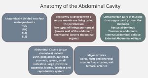
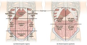
The abdomen can be divided into regions or quadrants. Understanding the structures within each quadrant or region helps healthcare professionals connect patient symptoms to a specific anatomical site and promotes clear communication (both verbal and written) as to the location of the symptom.
The abdominal cavity is the largest in the body; the outer layer is called the parietal peritoneum and is attached to the abdominal wall; the inner or visceral peritoneum envelops the internal organs in the respective peritoneal space. The diagram below shows that the liver, spleen, stomach, and intestines are enclosed in the visceral peritoneum, making them intraperitoneal. The kidneys and spleen are retroperitoneal, meaning they are not in the peritoneal space or surrounded by the peritoneal membrane (Thompson, 2018).
Abdominal Organs (Viscera)
Liver
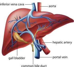
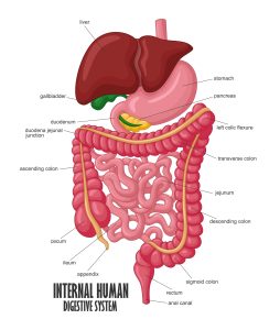
The liver is in the right upper quadrant of the abdomen, just below the diaphragm above the gallbladder on the right side of the abdominal cavity, and above the pancreas on the left. The liver contains four lobes and is highly vascular. The hepatic artery carries blood to the liver from the aorta. The portal vein transports blood from the digestive tract and spleen to the liver. The liver plays a major role in metabolizing carbohydrates, fats, and protein, producing substances required for blood coagulation, and storing many vitamins and iron. The liver acts as an excretory organ through bile production and converts fat-soluble waste to water-soluble material that the kidneys can excrete (Thompson, 2018).
Stomach
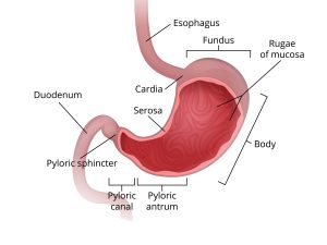
The stomach sits just below the diaphragm in the upper left quadrant and contains three sections: the fundus, the body, and the pylorus. The stomach secretes hydrochloric acid to break down fats and proteins (Thompson, 2018).
Gallbladder
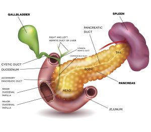
The gallbladder lies deep in the lower surface of the liver. Its principal function is to concentrate and store bile. Contraction of the gallbladder propels bile along the common bile duct to the duodenum, where it maintains the alkaline pH of the small intestine to make the absorption of lipids possible (Betts et al., 2022).
Pancreas
The pancreas is an exocrine gland that lies beneath the stomach. The pancreas produces digestive enzymes that break down proteins, fats, and carbohydrates. The pancreas also produces insulin and glucagon hormones (Thompson, 2018).
Spleen
The spleen is in the left upper quadrant, just below the diaphragm. The spleen is part of the reticuloendothelial system that stores and filters blood and manufactures lymphocytes and monocytes (Bickley, 2021).
Appendix
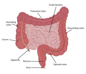
The appendix is located at the cecum, the beginning of the large intestine; its function is unclear, and newer research suggests it may serve as a secondary immune organ (Betts et al., 2022).
Small intestine
The small intestine is approximately 21 feet long; it begins at the pylorus. The first 12 inches of the small intestine make up the duodenum, and the final portion of the small intestine joins the large intestine at the ileocecal valve. The next eight feet of the small intestine is known as the jejunum; the ileum makes up the remaining 12 feet of the small intestine. Digestion occurs in the small intestine through the action of pancreatic enzymes, bile, and other enzymes (Thompson, 2018).
Large intestine
The large intestine begins at the cecum and terminates at the anus; it comprises four parts; the cecum and ascending colon, transverse colon, descending colon, and sigmoid colon. Most of the digestion of nutrients has already occurred in the small intestine; therefore, the primary function of the large intestine is to absorb the remaining water, vitamins, and electrolytes from waste material and produce and store solidified waste in the form of feces (Betts et al., 2022).
Kidneys and Bladder
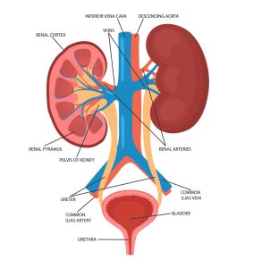
The kidneys are oval-shaped organs that sit on each side of the vertebrae at the level of T12 to T3. The right kidney sits 1-2 cm lower than the left kidney. The kidneys’ primary function is to excrete water-soluble waste in urine. The urine exits the kidneys and travels down the ureters to the bladder. The bladder is a hollow sac that serves as a reservoir for the urine released from the kidneys (Thompson, 2018).
Male and Female Reproductive Organs
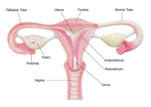
The female reproductive organs include the ovaries, fallopian tubes, uterus, cervix, vagina, and vulva (Bickley, 2021).
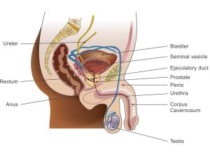
Male reproductive organs include the penis, scrotum, testicles, epididymis, Dusctus (vas) deferens, ejaculatory ducts, and urethra (Bickley, 2021).
Digestive System
Vasculature in the Abdominal Cavity
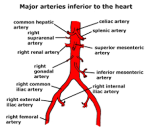
The aorta is the largest artery in the body; it descends from the left ventricle and bifurcates into the left and right iliac and femoral arteries. Before bifurcating, the aorta branches out to create arteries that supply blood to the liver, kidneys, spleen, stomach, and intestines (Betts et al., 2022).
